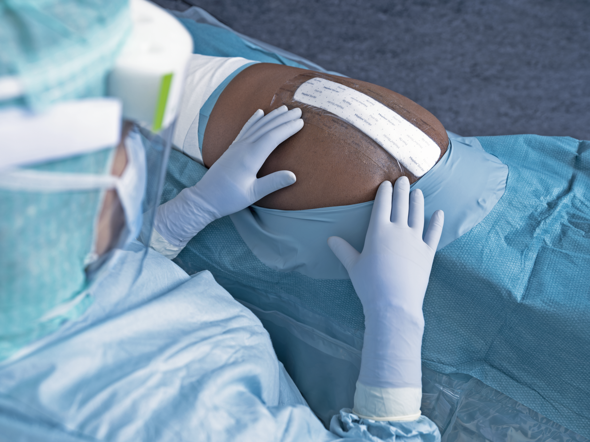
Pressure ulcer severity assessment is based on the International NPIAP/EPUAP pressure ulcer classification system and requires the clinician to determine the depth of the injury based on visual inspection of the wound and in the case of a stage 1 injury the presence or absence of non-blanching erythema. It should be noted that in most cases a pressure injury will develop over a bony prominence such as the heel or the sacrum (most common sites for pressure ulcers). Following initial staging of the pressure ulcer the clinician needs to ascertain the history of the wound in terms of how and when it was developed and the patient’s understanding of the wound and its causation. Note should be made of the degree of pain experienced by the patient related to the wound as well as any systemic signs of infection.
Staging
Category/stage 1
Non-blanchable Erythema. May be painful, firm, soft, warmer or cooler as compared to adjacent tissue.


Category/stage 2
Partial Thickness Skin Loss. Presenting as a shallow open ulcer with a red pink wound bed, without slough. May also present as an intact or open/ruptured serum-filled blister.

Category/stage 3
Full Thickness Skin Loss. Subcutaneous fat may be visible but bone, tendon or muscle are not exposed. Slough may be present but does not obscure the depth of tissue loss. May include undermining and tunnelling.

Category/stage 4
Full Thickness Tissue Loss. Exposed bone, tendon or muscle. Slough or eschar may bepresent on some parts of the wound bed. Often include undermining and tunnelling.

Suspected Deep Tissue
Depth unknown. Purple or maroon localised area of discolored intact skin or blood-filled blister due to damage of underlying soft tissue from pressure and/or shear. The area may be preceded by tissue that is painful, firm,mushy, boggy, warmer or cooler as compared to adjacent tissue.


Unstageable
Depth unknown. Full thickness tissue loss in which the base of the ulcer is covered by slough (yellow, tan, gray, green or brown) and/or eschar (tan, brown or black) in the wound bed.

Assessment for all stages
Location - Document anatomical location of PI
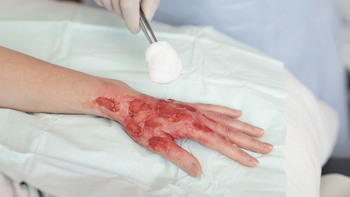
Tissue Type - Determine the characteristics of wound bed, noting the visible tissue which may include pink epithelisation tissue, red granulation tissue, yellow slough, black necrotic tissue
Exudate - Determine the type, amount and nature of wound exudate which may include, serous, serosanguinous, purulent and or viscous fluid
Odor - Note the presence of malodour from the wound
Wound Edge - Observe for undermining of the wound edges or any tunnelling which may be present
Peri wound - Describe the condition of the skin surrounding the wound and observe for maceration and inflammation
Infection - Assess for overt signs of wound infection
Document findings in individuals medical record (EMR)
* National Pressure Ulcer Advisory Panel, European Pressure Ulcer Advisory Panel and Pan Pacific Pressure Injury Alliance. Prevention and Treatment of Pressure Ulcers: Clinical Practice Guideline. Emily Haesler (Ed.). Cambridge Media: Osborne Park, Western Australia; 2014